Acetabular Dysplasia

What is acetabular dysplasia?

Acetabular dysplasia refers to an underdeveloped hip socket, or, the “acetabulum” the underdevelopment either affecting the bones, or the alignment/orientation of the socket or both. This bony problem, may in some people, result in increased movement (shearing) in hip joint resulting in instability (and in some cases dislocation) of the joint.  Early wear of the joint structures and ultimately hip pain and osteoarthritis¹ can eventuate.
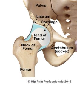
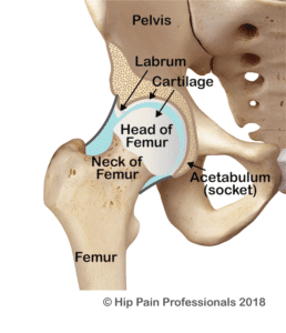
The hip socket is like a hemispherical cup that sits in the side of the pelvis. The opening of the cup faces out to the side and is normally tilted a little downwards and forwards. This provides a balance of joint mobility (flexibility) and stability. The socket and the ball (head of the femur) are normally of a similar shape and diameter, so that the socket hugs firmly around the ball (Figure 1). This allows the ball to swivel nicely around a centre point but controls excessive translation (sideways or back-forward motion). Optimal health of joint structures such as the cartilage (tissue that covers and protects the ends of long bones at the joints) and labrum (a triangular fibrocartilaginous fringe that joins the edge of the hip socket important for hip joint health) appear to be more easily maintained with this bony relationship between the ball and socket, that is, position, size and shape of the joints (outlined above) that allow for best mobility and stability.

Condition-Pages_Acet-Dysplasia_Fig1_Normal-258x300

Figure 1: Anatomy of the normal hip joint, with a good bony relationship. Cross-section through the middle of the joint.

Condition-Pages_Acet-Dysplasia_Fig-2_Type-1-258x300

There are two main variations to the shape of the hip socket, referred to as
“acetabular dysplasia”.

Types of Acetabular Dysplasia

1. Type 1: Sloping Roof Type (Figure 2)

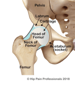
Type 1 acetabular dysplasia is where the opening of the hip socket is relatively wider than the ball.

The uppermost aspect (roof) of the socket slopes upwards, instead of curving around to hug tightly around the head of the femur.

This allows excessive “translation” or sliding of the ball in the socket which may place greater than normal loads on the joint structures (labrum, capsule, cartilage).

Condition-Pages_Acetabular-Dysplasia_Fig3_Type-2-258x300

2. Type 2: Short Roof Type (Figure 3)

In the second form of acetabular dysplasia, Type 2, the curvature and orientation of the cup is normal, but the roof of the cup is short.

This results in under-coverage of the upper aspect of the ball. This part of the ball and socket joint is the weightbearing aspect of the joint, so a smaller surface area will mean that when standing on one leg, the weight of the body is transmitted through a smaller area of cartilage.

Subsequently, this area of cartilage is exposed to relatively higher forces than normal. The labrum, which is the triangular piece of fibrous cartilage that sits around the rim of the socket (Figure 3) also becomes overloaded.

Due to the lack of bony support, the labrum ends up having to share some of the weightbearing force, which is not it’s normal role.

Diagnosis

Diagnosis is often missed in infants with routine checks. There are no clinical tests to diagnose this condition. The symptoms and clinical tests can be confused early on with other possible diagnoses and therefore clear diagnosis may take up to 5 years.  As acetabular dysplasia is a bony problem, medical imaging (usually an Xray) is required to make a firm diagnosis. Measurements can be made to determine the degree of dysplasia present. Magnetic Resonance Imaging may be used in addition to check if there has been any effect on the health of the joint cartilage and labrum.

What are the usual symptoms?

  • Pain is usually described as a sharp pain, felt deep at the front of the hip or groin, or around the front, outer corner of the hip.
  • Symptoms are often only present initially with more dynamic activities such as running, dancing or other sporting activities
  • Pain with activities that take the hip past 90 degrees up towards your chest, further aggravated if the leg is turned inwards.
  • Over time, symptoms may be present during everyday activities such as walking, getting in and out of the car, twisting when standing on one leg and climbing up or down stairs
  • It is also common to experience a feeling of weakness or heaviness of the leg, which is often described as a ‘dead leg’. With this feeling of weakness may come a lack of confidence in the stability of the leg, particularly when the weight is fully supported on that side, a feeling of “giving way”.
  • People with symptomatic dysplasia may also notice clicking and clunking noises coming from their hip. Lots of hips click however, so clicking alone does not mean there is a problem.
  • A waddle or a limp when walking may develop, associated with pain and/or muscle weakness

What causes acetabular dysplasia?

The exact causes of acetabular dysplasia are still not clearly understood.  The condition may be either present at birth or it may develop during childhood/adolescence, possibly linked to growth spurts or to ligament laxity/hypermobility. More research is required.

One-two percent of all babies are born with acetabular dysplasia². When a baby is born with this condition, it is usually referred to as either Developmental Dysplasia of the Hip (DDH) or congenital dysplasia of the hip. Congenital simply means something you are born with. In some newborns with very shallow sockets, the ball may easily slide out of the socket. In this case, a diagnosis of congenital dislocation of the hip may be given.

Condition-Specific-Pages_Acetabular-Dysplasia_Baby-in-Uterus-300x180
Condition-Specific-Page_Acetabular-Dysplasia_Swaddled-baby_iStock-184933720-1-200x300

Risk factors related to an increased likelihood of DDH include:

  • Being female: More than 80% of newborns with DDH are female³
  • Being first born.
  • Genetics: There may be a genetic factor running in the family, with likelihood increasing if there is a sibling with the condition, likelihood increasing further if one parent has the condition and even further if both parents have acetabular dysplasia4. There may also be a history of ligament laxity or hyper-mobility syndromes in the family.
  • Positioning of the baby in the uterus (womb): Likelihood of DDH is increased if the baby’s hips are held firmly crossed across the front of their tummy during pregnancy. This is more likely to occur if:
    • the baby is large and/or the uterus is small. As the uterus is usually smaller for the first pregnancy, the risk of DDH is greater for the first-born child
    • the baby has joint hypermobility (is extra-bendy)
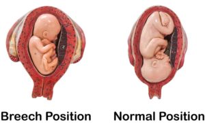
  • Positioning of the baby’s hips during birth: Likelihood of DDH is increased with a breech birth, where the baby passes through the birth canal bottom first, as opposed to head first.
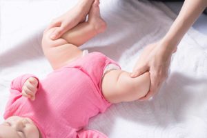
  • Positioning of the baby’s hips after birth: swaddling of babies, particularly of the legs is no longer recommended, because if the baby’s legs are held out straight and together for long periods, this can interfere with normal development of the hip sockets

In at least 40% of cases where DDH is diagnosed however, there are no identifiable
causes5

Condition-Specific-Page_Acetabular-Dysplasia_Frog-leg-baby-position_iStock-929515010-1-300x200

Getting Help. What is the treatment for Acetabular Dysplasia?

In most countries, new born babies are routinely screened for acetabular dysplasia.
A physical test is usually performed to assess for stability of the hip and if there is a. concern, an ultrasound assessment may be requested. If a diagnosis of acetabular dysplasia is made, excellent results can be achieved with double nappies, braces or splints to keep the hips in a ‘frog-leg’ type position.

This position allows the ball to push into the socket and help it mould around the ball shape of the head of the femur. Baby’s bones are still very elastic and change in shape can be achieved, which is why early diagnosis is best.

Many adults however function very well with mild acetabular dysplasia and are not even aware they have the condition. Acetabular dysplasia may only become a problem in situations where the alteration in bony shape is more severe, or if a lot of repetitive load is placed across a dysplastic joint (e.g. long-distance running).

If pain associated with acetabular dysplasia is diagnosed in an adult, the options depend on the age of the adult and the health of the joint. If there are already signs of hip joint osteoarthritis (cartilage wear), treatment for osteoarthritis will be offered. You can read more about osteoarthritis here.

Treatment for pain associated with acetabular dysplasia in a young adult with no osteoarthritis, includes:

Condition-Specific-Page_Acetabular-Dysplasia_Education_iStock-668440484-300x200

Treatment for pain associated with acetabular dysplasia in a young adult with no osteoarthritis, includes:

1. Education:

Understanding what the problem is and what types of positions and activities that are likely to irritate the hip joint, can help greatly in controlling the symptoms associated with acetabular dysplasia. Nutrition can play an important role.  Learning pain management techniques such as relaxation can be very useful.

Condition-Specific-Page_Acetabular-Dysplasia_US-lateral-Hip-300x218

2. Exercise:

Another key component of a treatment plan for painful acetabular dysplasia is a specific exercise program that includes:

a. Stability training

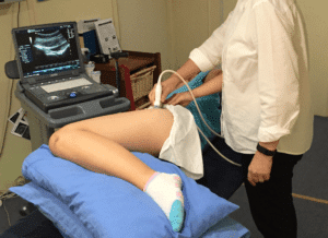
As the bony stability of the joint is reduced in acetabular dysplasia, specific exercises that target the deep cuff of muscles that hug around the hip can be useful. These muscles may help in reducing excessive translation (sliding) of the ball in the socket. Some health professionals, usually physiotherapists/physical therapists will use real-time ultrasound to assess and retrain these deep hip muscles. Ultrasound provides an excellent way to get real-time feedback on whether good activation in these muscles is being achieved.

Condition-Specific-Page_Acetabular-Dysplasia_Young-woman-limited-deadlift-with-light-bar_iStock-854374886-200x300

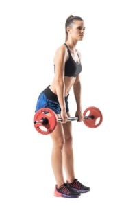
b. Strength training

In addition to good deep muscle support, general strength in the larger muscles around the hip and further down the leg can be helpful in controlling larger forces that cross the joint. For example, the large buttock, thigh and calf muscles can be very important in controlling landing forces absorbed up through the foot during walking, running and jumping. Good strength and endurance in these muscles will be important for controlling forces crossing the hip.

Condition-Specific-Page_Acetabular-Dysplasia_Gait_iStock-958225284-300x200

c. Movement training

The socket shape in acetabular dysplasia means that forces traveling through the hip will be absorbed over a smaller area or across a more sloping joint. In this circumstance, the direction of forces travelling across the joint is more important. The joint will be less able to control forces heading in directions where there is less than normal bony support. Particular attention to the way movement is achieved can be helpful in keeping the forces as much asb possible controlled within the main bony load-bearing areas of the joint. Some early research has shown that changing walking pattern can provide immediate and lasting improvements in pain and functional ability6.

3. Surgery

a. Periacetabular Osteotomy

For a small group of young adults, with more severe dysplasia and no osteoarthritis, reconstructive surgery may be offered as a treatment. This surgery is called Periacetabular Osteotomy and involves surgical restructuring of the bony hip socket. This is a very complex surgery with significant risks and should not be undertaken without good cause. If a surgical path is to be taken, a period of pre-habilitation can help promote a quicker recovery after surgery. Following surgery, there will usually be at least 3-6 months of rehabilitation to recover from the surgery, assist with adjusting to the new bone-muscle relationships and returning to everyday and sporting activities.

b. Hip arthroscopy

In cases of mild dysplasia, hip arthroscopy (keyhole surgery) is occasionally used to treat joint damage. However, the relative risks and benefits need to be carefully considered as the results are unpredictable and may lead to a worsening of symptoms7.

injection-therapy-350x250-1-300x200

4. Injections.

Injections into the hip joint

Hip injections into the joint using local anaesthetic and Corticosteroid (high dose anti-inflammatory) may be often used to help with diagnosis. If the injection into the joint helps improve symptoms, this is a reliable indicator that the pain is coming from within the hip joint itself.

These injections can also be used as a pain relieving method, sometimes helping to break the pain cycle so that physiotherapy / physical therapy can be commenced.

Always seek out a health professional with extensive experience and up-to-date knowledge in this area. Visit our directory to find a Hip Pain Professional near you.

References:
1. Harris-Hayes, M. and Royer, N. (2011). Relationship of Acetabular Dysplasia and Femoroacetabular
Impingement to Hip Osteoarthritis: A Focused Review. PM&R, 3(11), pp.1055-1067.e1.
2. Rosendahl, K. (1994). Ultrasound screening for developmental dysplasia of the hip in the neonate:
The effect on treatment rate and prevalence of late cases. Pediatrics, 94: pp.47-52.
3. Grill, F., Bensahel, H., Canadell, J., Dungl, P., Matasovic, T. and Vizkelety, T. (1988). The Pavlik
Harness in the Treatment of Congenital Dislocating Hip. Journal of Pediatric Orthopaedics, 8(1), pp.1-
8.
4. Wynne-Davies, R. (1970). Acetabular dysplasia and familial joint laxity: two etiological factors in
congenital dislocation of the hip. The Journal of Bone and Joint Surgery. British volume, 52-B(4),
pp.704-716.
5. Patel, H. (2001). Canadian Task Force on Preventative Health Care. Preventative health care, 2001
update: screening and management of developmental dysplasia of the hip in newborns. Canadian
Medical Association Journal, 164, pp.1669-77.
6. Lewis, C., Khuu, A. and Marinko, L. (2015). Postural correction reduces hip pain in adult with
acetabular dysplasia: A case report. Manual Therapy, 20(3), pp.508-512.
7. Parvizi, J., Bican, O., Bender, B., Mortazavi, S., Purtill, J., Erickson, J. and Peters, C. (2009).
Arthroscopy for Labral Tears in Patients with Developmental Dysplasia of the Hip: A Cautionary Note.
The Journal of Arthroplasty, 24(6), pp.110-113.

Condition-Specific-Page_Acetabular-Dysplasia_winning-shot_iStock-186546421-300x208

You might also be interested in these Blog Articles

cortisone-injections-for-trochanteric-bursitis-and-gluteal-tendinopathy

Cortisone Injections for Trochanteric Bursitis and Gluteal Tendinopathy

By hippainhelp | April 2, 2024

Pain at the side of the hip related to trochanteric […]

botox-injections-for-piriformis-syndrome

Botox Injections for Piriformis Syndrome

By hippainhelp | February 27, 2024

Have you heard of Botox injections for piriformis syndrome? Piriformis […]

exercises-for-gluteal-tendinopathy-and-hip-bursitis-common-mistakes

Exercises for Gluteal Tendinopathy and Hip Bursitis | Common mistakes

By hippainhelp | January 31, 2024

Which exercises should you avoid with pain at the side of your hip, gluteal tendinopathy and/or hip bursitis (trochanteric bursitis), or Greater Trochanteric Pain Syndrome (GTPS)

Recovering-from-Piriformis-Syndrome-Deep-Gluteal-Syndrome-or-Hip-Related-Sciatica

Sign Up To Our Mailing List!

For condition information you can trust, powerful self-help tips, myths, must-do's, don't do's and other helpful updates!

News Letter Page

Hidden

Next Steps: Sync an Email Add-On

To get the most out of your form, we suggest that you sync this form with an email add-on. To learn more about your email add-on options, visit the following page (https://www.gravityforms.com/the-8-best-email-plugins-for-wordpress-in-2020/). Important: Delete this tip before you publish the form.
Name(Required)
Are you a Health Professional? : tick box if YES