Hip Pain Explained will teach you about the anatomy of the hips and pelvic area and how many different types of body tissues interact. Each of these tissues is discussed in the tabs listed below: joint, soft tissue, bone, back referred, peripheral nerve and other. It is the interaction of these structures with the central nervous system and brain that result in the feeling of pain.
What is Pain?
Pain is an experience that the brain creates for the purposes of stimulating you to change your behaviour or seek help for a perceived problem with your body. Irritating or potentially damaging stimuli in your body (like high levels of pressure, tension or extremes of temperature) activate sense receptors (danger sensors) in the area. Signals from danger sensors in the body travel through the nervous system to the brain. Here the information is processed and the brain sometimes (but not always) produces a pain experience.
These danger sensors send warning signals to your brain, and your brain then decides whether the signals are dangerous to your health. Your brain uses lots of other stored and real-time information to make this decision, for example:
- whether you have had an injury to this area before
- whether you know someone who has had problematic pain or injury in this area
- whether an injury in this area is likely to impact substantially on your life (ability to work, play sport, stay healthy, interact with family and friends)
- what you have read or watched about the problem
- what others have said about the problem (friends, relatives, health professionals)
- if you are unwell with a cold or flu
- if you haven’t slept well
- if you are having a good or bad day
- if it’s raining or sunny, you are surrounded by harsh noise or calming music, pungent or pleasant smells …
After much scientific study, we now know that pain is not as simple as we once thought. It has been clearly demonstrated many times that:
degree of pain ≠ degree of damage.
Pain is rather a reflection of how dangerous the brain perceives the situation to be and how important it thinks it is for you to do something about it, based on subconscious considerations. The brain truly is amazing!
Persistent or cumulative signals from the danger sensors in your body, will usually trigger a pain experience, but at varying levels for each individual. Danger signals may come from many different structures around the hip and pelvis. The guide below serves as an introduction to these structures.
Pain experienced in the hip, groin and pelvic region may be related to:
- the joints of the hip & pelvis – hip joint, sacro-iliac joints, pubic symphysis
- the soft tissues – muscles, tendons, bursae & fascia
- bones – the femur (thigh bone) or bones of the pelvis
- local nerves running through and around the hip & pelvis
- musculoskeletal problems elsewhere in the body, such as the lower back (referred pain)
- problems with organs, other body systems or health issues masquerading as hip pain
Joint Related Pain
A joint is formed where two bones are joined together, with varying amounts of movement occurring between them. Pain may be related to the structures involved in the function and support of a joint.
There are several joints within the hip and pelvic region (Figures 1.1 and 1.2):
- the hip joints
- the pubic symphysis at the front of the pelvis
- the sacroiliac joints at the back of the pelvis
- the sacrococcygeal joint at the bottom of the tailbone (sacrum)
The Hip Joints
- The hip joint is the largest ball and socket joint in the body. The ball is the head of the femur (thigh bone). The socket in the pelvis, is called the acetabulum (Figure 1.3).
- Both the ball and socket are lined with smooth cartilage which allows the bones to slide against each other easily (Figure 1.3).
- The smooth cartilage lining the socket merges into a fringe of a more fibrous cartilage that joins around the edge of the bony socket. This is called the labrum (acetabular labrum) (Figure 1.3).
- It has a variety of functions, assisting in joint stability and health. The labrum makes the joint deeper and hugs firmly around the head of the femur, providing a suction effect. Both of these features contribute to joint stability. The labrum is also involved in the flow of nutrient-rich joint fluid and trapping fluid between the bones when you land on your foot in walking and running. This provides a cushioning effect for your cartilage and helps maintain joint health.
- The ball and socket joint is surrounded by a fibrous capsule, reinforced by a number of ligaments that run between the pelvis and femur (Figure 1.4). These strong ligaments provide stability for the joint but are also flexible, allowing your hip to move in large ranges of motion. There is a large normal variation in how flexible these ligaments are in different people.
Pain related to the Hip Joint
Changes in joint health:
- may occur in association with some conditions such as
- hip osteoarthritis
- femoroacetabular impingement syndrome (FAIS)
- acetabular dysplasia
- hip instability
- childhood developmental issues of the hip (Congenital dislocation of the hip, Perthes Disease, Slipped Upper Capital Femoral Epiphysis)
- DO NOT ALWAYS RESULT IN PAIN (Some level of wear or injury to tissues within the joint is so common in adults, it is thought to be normal. Identification of changes in joint health on XRays or scans is therefore not considered meaningful unless related to pain or other symptoms)
- are poorly correlated with level of pain
- most often become painful with one or more of the following factors:
- too much or too little activity
- rapid changes in activity levels
- activities that put large forces across the hip joint (e.g. stretching too far or landing heavily)
- certain sustained or repetitive postures or movement habits that create small changes that add up over time
- high body weight other general health factors
Pain related to the hip joint is most commonly experienced at the front of the hip (anterior hip), but may also be felt in the groin, side of the hip (lateral hip) or deep in the buttock. Other things may also cause pain in these areas, so visit our Pain Locator Map to read about different things that may be related to pain in each of these regions.
After a though assessment, your Hip Pain Professional will be able to:
- tell you whether the hip joint and changes on any XRays or scans, are likely to be related to your pain.
- assess for factors that could be contributing to the problem
- set a plan to help you:
- reduce your pain
- improve your ability to do everyday activities involved with work and the household
- return to sport or modified activity
- look after your hip joints for the long term, such as through targeted exercise and education around positions, stretches, exercises or activities that may not be ideal for your joint
- refer you for further investigation if required
- provide or refer you for further medical intervention, if appropriate (injections, surgical opinion)
The Pubic Symphysis
The pubic symphysis (Figure 1.5) is the joint where each half of the pelvis joins at the front of the body. The word ͚symphysis simply means – a place where two bones are closely joined. This joint is a flat or plane joint, that includes:
- a fibrous disc that sits between the pubic bones, working as a shock absorber
- four strong ligaments that support the joint
Only very small movements occur at this joint. For example, some rotation occurs during walking, as one side of the pelvis moves slightly forward and the other slightly back, following the movement of the legs.
Pain Related to the Pubic Symphysis
Pubic Symphysis pain is most commonly associated with injury or excess strain due to:
- a major trauma, such as a fall into the splits or a direct impact to the pubic region
- increased stretchiness of the joint ligaments during pregnancy – this is a normal response to hormone changes in preparation for childbirth
- trauma during childbirth – breech delivery, forceps/vacuum delivery
- a gradual build-up of joint stress, such as repetitive movements where the legs move far apart, as may occur in certain sports
- large repetitive forces created by the surrounding muscles, particularly the inner thigh muscles. This is most common in field or court sports that involve changing direction at high speed or kicking.
Pain related to the pubic symphysis is most commonly experienced in the groin region and/or directly over the joint. Groin pain that occurs over the pubic symphysis is referred to as Pubic Related Groin Pain. Other problems may also cause pain in the groin region, so visit our Pain Locator Map to read about different things that may be related to pain here.
Your Hip Pain Professional will assess your pubic pain and examine all the contributing factors. Advice and management approach will be determined by each individuals contributing factors – for example, previous trauma, pregnancy, level of conditioning and athletic involvement.
After a thorough assessment, a Hip Pain Professional will be able to tell you whether changes on your XRays or scans are likely to be related to your pain and what factors could be contributing to the problem. They will also set a plan to help you:
- reduce your pain
- improve your ability to do everyday activities involved with work and the household
- return to sport or modified activity
- look after your hip joints for the long term.
The Sacroiliac Joints
The sacroiliac joints (sometimes abbreviated to SIJs or SI joints) are the joints formed between the two bony wings of the pelvis and the tailbone (sacrum) (Figure 1.6).
- These joints are designed for stability with extremely little movement capacity. They are flat or plane joints though the surfaces of the joint are not completely flat. They are irregular or bumpy to allow some interlocking of the bones for the stability they need.
- They are also surrounded by some of the strongest ligaments in the body. This stable joint structure allows transfer of large forces between the body and the legs during walking, running and jumping.
- Some movement occurs in these joints during walking and running, where one leg is moving forward and the other backward, resulting in a small amount of joint rotation.
- Movement at the sacroiliac joints will also occur during extremes of hip or back motion, although most scientists agree that the amount of movement that occurs at the sacroiliac joints is very small.
Pain Related to the Sacroiliac Joint
Pain and functional difficulties related to the sacroiliac joint:
- most commonly develop during pregnancy or childbirth
- may occur after a major trauma, such as a hard fall onto the bottom or a large force through one leg, for example being dragged by one leg after a fall from a horse or water-skis.
- may develop over time, related to certain types of repetitive forces.
- can develop when there is a problem with the lower back or hip joints, transferring extra load across the sacroiliac joint.
Sacroiliac pain and dysfunction are however, greatly over-diagnosed. While true instability does occur, it is relatively rare and there are many people living unnecessarily in fear, related to a diagnosis of pelvic instability or being told their pelvis keeps moving out of place.
Here are a few facts about the sacroiliac joint that may dispel some of this fear:
- The structure of the sacroiliac joint makes it a very stable joint
- Asymmetry in the human body is normal
- Differences in tightness of the muscles that join on to either side of the pelvis occur naturally, related to leg or arm dominance, sport and occupation. This asymmetry may cause an appearance of asymmetry in the resting position and movement of the pelvis. This is normal and has not been linked to harm.
- Differences in leg length of up to around 1cm are common and normal. Leg length difference may also produce an appearance of asymmetry in pelvic position in standing.
- A click occurring in a joint does not mean it has moved ͚in or out of position. All of us click and pop, some just a little more than others. These are usually normal joint or tendon sounds.
There are also causes for sacroiliac joint pain that are related to other general health conditions. Sacroiliitis refers to an inflammation of the sacroiliac joints associated with a systemic inflammatory disease such as Ankylosing Spondylitis. You can read more about non-musculoskeletal causes of hip and pelvic pain here.
Pain related to the sacroiliac joints is most commonly experienced in the upper buttock region, usually right over the joint, in the area of the dimples at the top of the buttocks. As the pelvis is a ring joined at the front by the pubic symphysis, problems with the sacroiliac joints are sometimes associated with pain in the groin region. There are many other causes for groin pain however, so visit our Pain Locator Map to read about different factors that may be related to pain in each of these regions.
Your Hip Pain Professional will be able to assess your sacroiliac joints to determine if they are likely to be the cause of your pain. If you have sacroiliac joint related pain, you may require:
- advice on modifications to activities or positions – e.g. workplace set-up
- an exercise program to provide optimal muscle support around the joints
- advice regarding short term bracing or taping – only appropriate for short term use and usually only for severe pain and during late stage pregnancy or after trauma
- an injection for short term relief of severe pain, while you work on your rehabilitation
The Sacrococcygeal Joint
- The sacrococcygeal joint is the joint in the tailbone formed between the sacrum and the coccyx (Figure 1.7).
- The coccyx is a small triangular shaped bone made up of 3-5 fused segments. Many ligaments attach to the coccyx helping to provide stability and support for the pelvis, its muscles and contents.
Pain related to the Sacrococcygeal Joint – Coccydynia
Pain relating to the coccyx and sacrococcygeal joint is known as coccydynia (pronounced cox-y-dynia). A sudden onset of coccygeal pain is usually associated with trauma, externally such as from a fall backwards onto the bone, or internally such as in childbirth. Onset can also be gradual, commonly related to sustained pressure from sitting or local muscle forces.
Factors associated with an increased risk of developing coccydynia are:
- being overweight
- being female – women are 5 times more likely to develop the condition
- increased flexibility or mobility of the joint (hypermobility)
- reduced flexibility or stiffness of the joint (hypomobility)
- variants of coccyx shape
- pelvic floor muscle weakness or overactivity
- other general health factors (see other causes section): These must be ruled out.
Your Hip Pain Professional can reduce your pain associated with coccydynia by addressing factors such as:
- excessive weight bearing on the coccyx due to seating or habitual sitting postures
- weakness, spasm or altered activity in the muscles that attach to the coccyx including the gluteus maximus (buttock) and pelvic floor muscles (see the soft tissue section)
- altered stress and strain on the structures that attach to the coccyx which may have occurred after trauma or as a gradual build up over time.
- providing or referring you for a pain-relieving injection into the area or in very rare cases, to a surgeon.
Soft Tissue Related Pain
Pain around the hip and pelvis may also be related to soft tissues (Figure 2.1). Soft tissues are non-bony structures that connect, support, or surround other structures and include:
- the muscles, which allow you to move
- the tendons, which connect your muscles to the bone
- the bursae – small, flat pockets of fluid that help all these things slide and glide against one another, reducing friction. There are bursae are all around the body in places where different structures may rub against one another, for example where a tendon runs around a bone.
- the fascia – stretchy, thin, white fibrous tissue. All our muscles are enveloped in fascia, like stretchy stockings that help transfer energy from muscle and movement. Fascia also forms sheaths or tunnels for safe passage of blood vessels and nerves and wraps and supports all our bodily organs.
There are many muscles that support and move the hip and pelvis. The soft tissues located in each of the main areas of our hip and pelvis will be discussed below. This information can also be found by selecting your specific area of pain or difficulty on our Pain Locator Map.
These different areas are:
Soft Tissues of the Anterior Hip Region (Front):
The muscles that sit at the front of the hip are called the hip flexors (Figure 2.2) and act to lift your knee towards your chest (flexion). The main hip flexor, the iliopsoas muscle is made up of two parts – the psoas muscle that starts at the lumbar spine, and the iliacus that starts from the inside of the pelvis.
They come together in the pelvis and run forwards over the front of the pelvis, deep across the front of the hip and join onto the upper thigh bone (femur). The iliopsoas muscle provides important support for the front of the hip joint, the sacroiliac joint of the pelvis and the lower back (lumbar spine). It is also a powerhouse for moving your legs forward in walking, running and stairclimbing.
A number of other longer, more superficial (closer to the surface) hip flexor muscles run from the pelvis and down into the thigh and connect below the knee – the Sartorius, Rectus Femoris and Tensor Fascia Lata (TFL) muscles (Figure 2.3). These muscles help the iliopsoas muscle with lifting the thigh, but their effect also extends across the knee.
Hip Flexor or Iliopsoas Related Pain
Tears or strains may occur within the hip flexor muscles; where the muscles and tendons join (musculotendinous junction); or within the tendons. Traumatic injury resulting in rapid onset of pain is usually associated with sporting actions such as:
- kicking
- sprinting, particularly rapid acceleration
- changing direction at speed.
Changes in tendon health often evolve more slowly, related to your bodys natural make-up (e.g. bony shape, hypermobility) or postural, movement or athletic training habits. The iliopsoas tendon that joins the iliopsoas muscle to the femur (thigh bone) sits deep at the front of the hip and is separated from the hip joint by a large bursa (iliopectineal or iliopsoas bursa). This bursa reduces friction and helps the tendon slide freely across the front of the hip.
The tendon and/or the bursa may become involved and you may receive a diagnosis of:
- iliopsoas tendinopathy – a painful iliopsoas tendon
- anterior snapping hip – snapping of the iliopsoas tendon which can sometimes become painful
- iliopsoas bursitis – inflammation or thickening of the iliopsoas bursa
- iliopsoas impingement – impingement or compression of the iliopsoas tendon against the underlying bone
The hip flexors muscles, and particularly the iliopsoas muscle, have become incorrectly demonised in the popular media for being the source of many problems. These are generally thought to be related to the muscles becoming overly active and tight. There is unfortunately much misinformation on the internet regarding the hip flexors and poor advice regarding management of true hip flexor issues.
Many people are stretching the hip flexors and placing high loads on the front of the hip unnecessarily, often worsening their problem.
Your Hip Pain Professional can:
- assess the hip flexors and decide whether stretching will help or aggravate the problem
- provide important advice about sporting activities, for example:
- advice re how much is too much
- allowing adequate recovery between sessions
- temporary reduction in activity levels may be required
- rest from particular actions that place highest load across the hip flexors and pelvis
- provide or send you for rehabilitation to optimise function of these muscles
Soft Tissues of the Groin Region:
The adductor muscles of the groin sit on the inner side of the thigh, between the pelvis and the knee (Figure 2.4). This group of muscles includes:
- the adductor magnus (large adductor)
- the adductor longus (long adductor)
- the adductor brevis (short adductor), that sits underneath the adductor longus muscle
- the gracilis (a long strappy muscle)
- pectineus (a smaller muscle)
These muscles can be involved in other roles but primarily work to pull the legs inwards, towards each other (adduction). They also help control the energy of the moving leg or the moving body during kicking actions and changing direction when running. As the body moves away from the planted foot when ͚cutting͛or dodging, the inner thigh muscles have to control the momentum of the moving body.
The adductor longus and often the brevis too, have extensive connections through the groin (Figure 2.5), into:
- the abdominal muscles at the front of the trunk
- the inner thigh (adductor) muscles of the opposite side
- the joint at the front of the pelvis (pubic symphysis) and the blend of fibrous tissue that runs across this joint (the pubic aponeurosis)
The abdominal muscles extend from the ribcage down to the pelvis, supporting the spine and allow the trunk to curl upforwards or to the side. They also help control the position of the pelvis. The rectus abdominis (the ͚6 pack͛ muscle) joins onto the pubic bone and connects into the adductor muscles and the pubic aponeurosis (Figure 2.5). There are also another 3 layers of abdominal muscles (external oblique superficially, internal oblique beneath and
Figure 2.6: The soft tissues of the abdomen and hips as viewed from the front. Area of inguinal related groin pain is indicated by the dashed box. transversus abdominis deepest) that wrap around your body like a corset. At the front, they join into a big ligament that runs across the groin – the inguinal ligament (Figure 2.5).
Adductor Related Groin Pain
Tears or strains may occur within the adductor muscles; where the muscles & tendons join (musculotendinous junctions); or within the tendons. Traumatic injury resulting in a rapid onset of pain, is usually associated with sporting actions such as:
- changing direction at speed
- sliding sideways
- kicking
A single cause for more longstanding groin pain can however be much more difficult to establish. One of the main reasons for this is the large amount of interconnection between the soft tissues around the pubic region. Magnetic Resonance Imaging (MRI) scans of someone who has had groin pain for more than 3 months will often reveal a variety or combination of findings, and you may receive a diagnosis of:
- adductor tendinopathy, tendinitis or tear – tendon pain, inflammation or a tear of one or more of the inner thigh muscles
- pubic aponeurosis tear – a tear in the blended fibrous tissue at the front of the pubic bone
- rectus abdominis tendinopathy/enthesopathy, tendinitis or tear – tendon pain, inflammation or a tear of the rectus abdominis (six pack) muscle
Inguinal Related Groin Pain
In the region where the abdominal muscles join onto the pelvis and the inguinal ligament (Figure 2.6), tears or weakening of some of the connections can occur, leading to pain and/or a hernia. A hernia is where the pressure of the bowel against the weakened area can cause the appearance of a bump, as the bowel pushes into the area. More severe hernias are easily visible, but most of the time inguinal hernias are small and only picked up on ultrasound scans.
Your Hip Pain Professional can:
- provide a thorough assessment of all the soft tissues in the groin region
- refer you for appropriate scans, if necessary
- provide or refer you for rehabilitation of this area,
- provide advice and/conditioning for successful return to sport
- refer you to a surgeon or provide surgery (if your HPP is a surgeon) – this is rarely required but may be necessary with more severe hernias or abdominal tendon tears.
Soft Tissues of the Lateral Hip Region (Side):
The muscles that sit around the side of the hip are called the hip abductor muscles (Figures 2.7 and 2.8). Their main functions are to move your leg out away from the midline of your body (abduction) and to anchor your pelvis to your femur when standing on one leg. This allows you to keep your pelvis fairly level and maintain your balance.
There are 3 main layers of hip abductor muscles:
- 1. Deepest layer – gluteus minimus (smallest muscle), which joins into the hip joint capsule and is thought to have an important role in supporting this joint. It runs from the outer side of the pelvis to its anchor-point on the femur (greater trochanter) (Figure 2.7).
- 2. Middle layer – gluteus medius (middle sized muscle), which also joins the pelvis to the top of the femur (greater trochanter) and is one of the main muscles that helps control pelvic position (Figure 2.7).
- 3. Superficial layer – the upper part of the gluteus maximus (largest gluteal muscle) and the tensor fascia lata (TFL) muscle. These muscles create their effect at the lateral hip through their connections to a long fibrous band that runs down the outside of the hip, thigh and knee, called the iliotibial band (ITB) (Figure 2.8).
With all these muscles and their tendons moving over each other and the underlying bones, a number of small flat, fluid-filled cushions (bursae) are present, to help everything slide freely (Figure 2.7). The main bursae are the trochanteric bursa, the subgluteus medius bursa and the subgluteus minimus bursa.
Lateral Hip Pain
Acute muscle tears or strains occur most frequently in the gluteus maximus and medius muscles and occasionally in the tensor fascia lata muscle. This is more likely to occur during rapid sidestepping or change of direction when running. Tears in these muscles are however relatively rare compared to thigh and groin strains.
Pain over the side of the hip that develops more gradually is most commonly related to the health of the soft tissues at the anchor-point for the gluteus medius and minus on the femur (the greater trochanter) (Figure 2.7). You may receive a diagnosis of:
- Gluteal tendinopathy, tendinitis or tear – tendon pain, inflammation or a tear of one or more of the gluteal tendons
- Gluteus medius or minimus tendinopathy, tendinitis or tear – tendon pain, inflammation or a tear of the gluteus medius of minimus tendons
- Trochanteric bursitis, hip bursitis or even just bursitis – an irritated bursa
- Greater Trochanteric Pain Syndrome (GTPS) – a combination of some of the above
You might like to read further about terms used for lateral hip pain in our blog.
Pain in this area is often attributed to tightness of the ITB and therefore you may read much advice to stretch the ITB as a self-help strategy.
There is however NO evidence to support stretching the ITB as a self-help strategy. In fact, expert Hip Pain Professionals agree that people with gluteal tendon or bursal problems are much more likely to be longer than average, rather than short in the ITB. Stretching may actually provoke the pain, rather than settle it (Figure 2.9).
Read more here on the high-quality scientific evidence that is now available on successful management of this condition (with NO ITB stretching!).
Seek the assistance of a Hip Pain Professional to:
- test if your hip abductors are short & tight or actually too long – many people who have poor control of their hip, especially in single leg weight bearing activities, actually drop their hip out too far, their muscles feeling tight as they work overtime to try to provide support. Stretching will not help and may make things worse.
- work out if your hip abductors have any part to play in your problem and provide a plan for recovery
A Hip Pain Professional can:
- test if your ITB or hip abductors are tight (shortened) or not, and provide guidance as to whether stretching will help or aggravate the problem
- provide simple advice for controlling aggravating positions and actions that can immediately help you to start getting your pain under control
- provide or direct you towards a high-quality rehabilitation program that has been shown to be successful under rigorous scientific conditions
- provide advice regarding the use of steroids or other injections in cases of severe pain or pain unresponsive to evidence-based rehabilitation.
Soft Tissues of the Upper Buttock Region:
In the upper buttock region, many muscles and their surrounding thin fibrous wrapping (fascia) converge, joining firmly onto the pelvis and sacrum. These back, abdominal and buttock muscles play a vital role in generating and transferring forces that pass across the pelvis, between the back and hips. The muscles are also covered in strong fascia that continues from the hips (gluteal fascia) into the back (thoracodorsal fascia). This strong, stretchy wrapping helps store and transfer energy through this region (Figure 2.10).
The upper part of the gluteus maximus muscle, and the gluteus medius muscle beneath, run from their anchor points on the pelvis and sacrum, around to the side of the hip (lateral hip region). The piriformis muscle also sits under the gluteus maximus in the upper buttock, having strong connections into the bones and ligaments of the sacroiliac joint (SIJ) at the back of the pelvis (Figure 2.11)
Soft Tissue-Related Pain in the Upper Buttock Pain in the upper buttock can be associated with any of the local soft tissues – the muscles, their attachments onto the bone and the fibrous wrapping (fascia) that surrounds them.
Problems in these soft tissues may develop due to:
- an acute injury, usually related to heavy lifting or twisting or a sports injury
- weakness in the buttock or trunk muscles
- overuse of the buttock or trunk muscles. Increasing bulk in muscles that are working too hard also has the potential to worsen pain.
- general overload – doing too much of a particular task or activity; increasing activity too quickly, without adequate recovery
- general underload – a sedentary lifestyle with too little stimulus to keep yourmuscles healthy. This may also happen after a period of relative rest, for example after travelling, or after being ill.
A Hip Pain Professional can:
- assess the buttock and trunk muscles that may contribute to pain in this region
- determine if a strengthening program is needed, which exercises are likely to be most effective and which are best to avoid
- assess if there is excessive activity or gripping of the muscles in this area and provide strategies in how to combat this
- provide important advice about sporting and other activities
- advice about ͚how much is too much
- allowing adequate recovery between sessions
- temporary reduction in activity levels may be required
- rest from particular actions or altering techniques that place high stress through the upper buttock region
Soft Tissues of the Lower Buttock Region:
The muscles of the lower buttock region are involved in providing support for the back of the hip joint and moving the hip into extension and external rotation – taking your leg backwards and turning your knee outwards.
If you put your hands on the lower half of your bottom you will be touching the lower portion of the gluteus maximus muscle, an important muscle for pushing up from a squat or lunge position or for extending the leg behind especially when walking up a hill or stairs (Figure 2.12).
The hamstring muscles (semimembranosis, semitendinosis and biceps femoris muscles) in the back of the thigh help to extend the hip (take the thigh backwards), but also bend the knee. This combined function makes the hamstring muscles very important muscles for transferring forces between the hip and lower leg in actions such as running, kicking, lifting and lunging (Figure 2.12).
Beneath the lower part of the gluteus maximus muscle, sit a group of small muscles, referred to as the deep hip rotators (Figure 2.13). They work to rotate the thigh and turn the knee outwards. They also have important connections into:
- the back of the hip joint, allowing them to provide extra stability for the hip joint
- the pelvic floor muscles.
The hamstring muscles in the back of the thigh also attach deep in the buttock, onto the sitting bones (ischial tuberosities). Directly on top of the hamstring attachment is a small flat, fluid-filled cushion (the ischial or ischiogluteal bursa) (Figure 2.13). This helps reduce friction and allows smooth gliding of the largest buttock muscle (gluteus maximus) over the hamstring tendons.
Soft Tissue-Related Pain in the Lower Buttock Region:
Soft tissue related pain can occur in the Lower Buttock Region for many reasons:
- Muscle soreness, tears or strains may occur in this region related to large or unusual forces across the largest buttock muscle (gluteus maximus) or the deep rotator muscles. Tears of the deep rotator muscles may occur with rapid change of direction or a twisting injury.
- The deep rotators may also become overactive and painful associated with weakness or excessive activation of other muscles in this area (including the pelvic floor, gluteal or hamstring muscles).
- Ischiofemoral impingement is a diagnosis referring to a situation where one of the deep external rotator muscles (quadratus femoris) is repetitively compressed in a smaller than normal gap between the sitting bone (ischial tuberosity) and the thigh bone (femur).
- Hamstring tendon pain may come on quickly, in response to a large strain, often a slip or fall, or a rapid change in activity. However, changes in tendon health often evolve more slowly, related to postural, movement or athletic training habits, particularly long distance and hill running.
- Ischial or ischiogluteal bursitis is a diagnosis referring to inflammation of the small, flat, fluid-filled cushion called the ischiogluteal bursa (Figure 2.13) that sits over the hamstring tendons at the sitting bone. The bursa may become inflamed and painful when it is struggling to cope with high levels of compression or friction over the sitting bone, possibly associated with sitting on hard surfaces or athletic activity such as running uphill.
Your Hip Pain Professional can:
- assess the gluteus maximus and deep hip rotator muscles
- assess the hamstring muscles and their tendons
- provide guidance as to what exercises will be most beneficial, for example hamstring stretching is usually not helpful for those with hamstring tendinopathy or tendon tears.
- assess if weakness is an issue and if so, exactly which of the muscles are weak or activating too much, thus providing you with exercises specific to your individual needs
- assess if ‘neuromuscular’ control is an issue, that is, do you move in a way which may irritate or overload the painful structures
- provide important advice about sporting activities, such as
- ‘how much is too much’
- allowing adequate recovery between sessions
- altering activity levels to match soft tissue tolerance
- resting from or altering particular actions that place highest load across the lower buttock region
- provide advice on seating if pain while sitting is an issue
- advise if injections or surgery may be warranted
Soft Tissues of the Saddle Region:
The saddle region is the area between the hips that would contact a saddle when riding a horse or bicycle. There are a number of muscles and large ligaments that run through this region.
The pelvic floor is the name given to a group of muscles that form a wide sling between the pubic bones at the front of the pelvis and the lower sacrum and coccyx (tailbone) at the back (Figure 2.14 and 2.15).
These muscles (levator ani and coccygeus), have a number of important functions:
- to support the organs of the abdomen and pelvis (e.g. bladder, bowel, uterus)
- to resist increases in pressure in the abdomen (intra-abdominal pressure), for example, coughing, laughing and lifting.
- urinary and faecal control (continence)
The pelvic floor muscles have strong connections with one of the deep hip rotator muscles (obturator internus). This muscle starts from the inner surface of the pelvis in the saddle region, then runs out through the back of the pelvis and across the back of the hip, through the lower buttock region (Figure 2.15). You can read more about the deep rotators in the section on Soft Tissues of the Lower Buttock.
There are some large ligaments (Figure 2.16), the sacrotuberous and sacrospinous ligaments, that sit within the pelvis and connect between the sacrum and the pelvis. The sacrotuberous ligament runs from the sacrum to the inner edge of the ischial tuberosity (sitting bone) where it has connections into the hamstring tendons.
The sacrospinous ligament runs from the sacrum, underneath the sacrotuberous ligament and across to an area above the sitting bone, called the ischial spine. The sacrospinous ligament has strong attachments to the coccygeus muscle of the pelvic floor. These ligaments play an important role in providing stability for the sacroiliac joints and assisting the pelvic floor in supporting the internal organs.
Soft Tissue-Related Pain in the Saddle Region
Pain in the saddle region may be due to problems with the pelvic floor muscles, often referred to as pelvic floor dysfunction. The muscles may be too weak or too active. If they are too active, known as a hypertonic pelvic floor then strengthening exercises may not be the best approach. Pelvic floor dysfunction may result in:
- leakage or incontinence, urinary or faecal
- difficulty with passing urine, or moving the bowels
- pain: this can be in the saddle area but may also be felt in the groin, buttock or lower back.
Vaginismus is a diagnosis where the hypertonic pelvic floor (pelvic floor muscles that are too active) or muscle spasm affects the muscles around the vagina, making any penetration painful or impossible, for example, inserting tampons or sexual intercourse. This is a complex condition and can have many causes. Some medical causes need to be ruled out. Psychological issues can also be relevant and must be addressed. However, in cases linked in particular to trauma (often from childbirth) or micro damage occurring around menopause, musculoskeletal therapy can be beneficial.
Coccydynia is a diagnosis meaning a painful tailbone. This may develop in association with either weakness or overactivity/spasm in the pelvic floor muscles. (You can also read more about pain relating to the coccyx and sacrococcygeal joint, known as coccydynia, under the joint section of Hip Pain Explained)
Pain may also be associated with abnormal strain placed on the large ligaments (Figure 2.16) of the pelvis (sacrotuberous or sacrospinous ligaments). This is usually secondary to problems with the sacroiliac joint, hamstrings, or pelvic floor muscles.
Your Hip Pain Professional can:
- assess the hip and pelvic region to identify problems that may be influencing the health of the soft tissues of the saddle region
- determine if some form of assessment of the pelvic floor muscles is required. This may involve:
- non-invasive ultrasound assessment of pelvic floor function
- an internal assessment from a Womens or Mens Health physiotherapist, a gynaecologist or pain specialist with a special interest in this area or your general practitioner. This can give much more information on the type of muscle difficulties (weakness or excessive activity (hypertonicity)), the severity of the difficulty and an indication of which course of treatment is likely to be most effective.
- provide a variety of options for management of your particular type of problem. This may involve:
- strategies to either strengthen or relax your pelvic floor muscles
- self-massage or massage provided by a health professional trained in these techniques
- home exercises or muscle relaxation strategies
- other brain-based techniques (mindfulness, meditation, visualisation etc) or counselling to address any issues underlying pelvic floor muscle spasm or pain.
Bone Related Pain
Although relatively more rare than soft tissue or joint problems, bony problems such as a fracture, stress fracture or very rarely, infection or tumour within the bone may cause hip and pelvic pain.
In growing children or adolescents, pain may be related to disorders of the growth plates of the femurs or pelvis.
The Bones of the Hip & Pelvis
The pelvis is a ring of bone, made up of two halves, joining together at the front in the pubic symphysis. At the back of the pelvis, these halves join to either side of the sacrum which is the lowest part of the spine. These joins form the two sacroiliac joints (SIJs).
Beneath your sacrum is your coccyx, joining at the sacrococcygeal joint. These are the bones that make up the pelvis(Figures 3.1and 3.2).
More information about these joints can be found under the Joint tab listed above.The long bone within the thigh is called the femur. It has a ball on the top called the head of the femur. This fits into a socket on the pelvis called the acetabulum. This is the hip joint (Figure 3.1)
Bone Related Pain
Bone related pain may be due to:
- Fracture
- Stress fracture
- Avulsion fracture
- Apophysitis
A fracture:
- is the medical term for a broken bone
- usually occurs from a traumatic event like a fall, car accident or more severe sporting injury
- is more likely to occur in a weakened bone e.g. osteoporosis or genetic diseases like osteogenesis imperfecta
The most common fractures around the hip and pelvis include a fracture of the neck of the femur (common in older women with osteoporosis), or fractures through the pelvis related to large traumas or where stress fractures have not been attended to early enough.
Sometimes the coccyx (tailbone) may be fractured during childbirth or a heavy fall onto the bottom or back may result in a fracture of the coccyx or sacrum.
A stress fracture:
- is the medical term used for a fracture that occurs due to a build-up of repetitive stress
- is usually related to overuse – an accumulation of repeated small traumas e.g. long-distance running
- is more likely to occur with rapid increases in athletic training volume and/or inadequate recovery time
- starts as localised swelling in the area of bone exposed to highest stress (you won’t see this at skin level as it is just within the bone) – this may be called a stress reaction or bony stress response
- may progress to full fracture if you don’t rest Stress fractures may occur in a number of sites around the hip and pelvis (Figure 3.3), most commonly the top of the thighbone (neck of the femur), the bones at the front of the pelvis (the pubic ramus) or the tailbone (sacrum)
An avulsion fracture:
- is where a small piece of bone is pulled away from the main part of the bone
- may occur in adults where a large force across a ligament (joins bone –bone) ortendon (joins muscle to bone), causes a small piece of bone to come away with the ligament or tendon
- may occur in children at the bony growth plates (read more in the next section)
Apophysitis:
- usually referred to as ‘growing pains’
- occurs in adolescents
- is due to long bones such as the femur growing more quickly than the muscles, which then tug on the growth zones in the immature pelvis
- most commonly occurs at sites where some of the large muscles attach:
– at the front of the pelvis
a) where the sartorius muscle joins to the ASIS (anterior superior iliac spine) or
b) where the rectus femoris muscle joins to the AIIS (anterior inferior iliac spine)
– at the back of the pelvis where the hamstring muscles attach to the sitting bone (ischial tuberosity) in the lower buttock (Figure 3.3).
(You can visit the ‘Soft Tissue Related’ tab to view these muscles)
The pain of apophysitis is related to an inflammation of the bony growth centres where these muscles attach. Pain is generally experienced locally at the area of muscle attachment but can radiate around the area as well.
Other growth-related bony issues:
- Perthes disease or Legg-Calvé-Perthes Disease – is misshaping of the femoral head related to issues with blood flow to the bone. Most common during ages 4-10 years.
- Slipped Capital Femoral Epiphysis or Slipped Upper Femoral Epiphysis-usually a small shift in the growth plate in the neck of the femur that can change the shape of the head and neck of the femur. Most common during ages 8-15 years.
Pain related to the bones could be felt anywhere around the hip or pelvis region depending on the bone affected and the cause of the bone pain. Our Pain Locator Map highlights the bones that may be responsible for pain in each region.
Pain in these areas may have other causes, so visit our Pain Locator Map to read further.
Your Hip Pain Professional can:
- perform a thorough assessment and let you know if a bone issue may be suspected. In these cases, you may be advised to undertake further imaging or referral to a medical specialist for further advice
- provide or refer you for rehabilitation at an appropriate time following bony injury
- provide important information about managing sporting activity and recovery in those with bony stress injuries or adolescents with bony ‘growing pains’
- address muscular or bio mechanical issues that may be contributing to bone-related pain, for example, running style may have an impact.
Back Related Hip Pain
Pain experienced around the hip and pelvis sometimes has nothing to do with problems in this area.
Problems in the lower back can result in back pain and/or pain through the hips and down into the legs. There are two main ways this might occur:
Referred pain
Referred pain is pain felt in a part of the body other than its actual source. For example, if there is a problem in the lumbar (lower back) discs or joints, small nerve endings serving these structures generate ‘danger’ messages that are transmitted along small nerve fibres into the spinal cord.
However, this area of the spinal cord also receives information from structures in the hip and pelvis.The brain is unable to distinguish where the information came from, (the back, the hip or the pelvis) so you might feel pain in any one or a combination of these areas (Figure 4.1).
Referred pain:
- is usually a dull, aching or gnawing pain
- can expand into a wide area that is difficult to localize
- is not related to a problem of the nerve roots in the spine
- is not associated with other nerve-related symptoms such as tingling or numbness
Radicular Pain
Radicular pain is pain associated with irritation of the nerve roots as they exit the spine. Most commonly this is associated with inflammation or compression from the nearby disc. The nerve roots that exit at each level of the spine give rise to sensation indifferent areas of the skin, called dermatomes (Figure 4.2).
When a nerve root is irritated at the spine, pain may be felt in the area of skin that the nerve root supplies. For example, compression of the nerve roots that exit between the 4th and 5th lumbar vertebrae (L4-5) or the 5th lumbar vertebra and the sacrum (L5-S1),could, for some people, result in painful sensations across the buttock, down the back of the thigh and right down into the foot (see picture).
The nerve root compression of the lower levels (L4, L5 and S1 and their resulting referred pain is often called “sciatica”.
This term is not correct. The Sciatic Nerve is formed where the L4-S1 nerve roots blend together outside of the spine: the term “sciatica” refers to irritation of this nerve, NOT the nerve root.
So where the irritation is at the nerve root, before the nerve roots actually blend together to form the sciatic nerve, it is incorrect to use the term “sciatica.”
The term “radicular pain”is now used instead.
Radicular pain:
- is related to a problem of the nerve roots in the spine
- is usually accompanied by stabbing or shooting pains
- is usually easier to localise than referred pain
- may also be associated with an additional or background, deep, dull ache
- may be accompanied by other nerve related symptoms such as tingling, itching, burning or numbness
If the pain in your hip is due to a problem in the back, you can spend valuable time and money getting unnecessary treatment on the wrong area and this might delay an important diagnosis and your recovery.
It is also important to be aware that pain in some regions, particularly lateral hip and buttock pain, is often assumed to be back-related pain when the pain may be all or partly due to a problem of the hip.
Again, you may spend valuable time and money getting unnecessary or incomplete treatment.
Your Hip Pain Professional can:
- provide a skilled assessment
- help figure out the actual source of the problem and if you have more than one area contributing to the problem
- develop a comprehensive plan to help the problem – If your pain is lumbar-referred or radicular, you may need treatment on your back and not your hip or if you have two problem areas, both will need to be addressed within a comprehensive management plan
- recommend further tests or refer you to another specialist if the problem does not appear to be in the musculoskeletal system.
Peripheral Nerve Related Pain
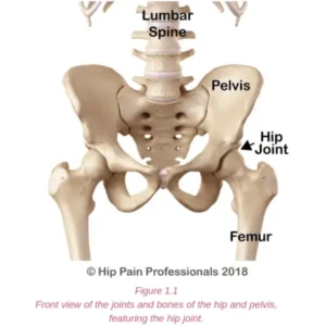
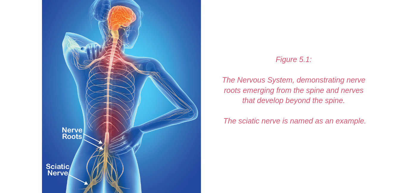
The nervous system (Figure 5.1) is a complex network of nerves and cells that carry messages between the brain and spinal cord and your body. It is through this system that we feel, move and control our bodily functions.
Nerve roots leave the spinal cord via the intervertebral foramina (holes or spaces between the vertebrae) and join together from various levels of the spine to travel as cord-like structures, called nerves, to their destinations.
It is these nerves that travel outside the spinal cord that are referred to as “peripheral nerves”.
Some peripheral nerves travel only a short distance and others all the way from the lower back to the foot. Along their journey they run between and through muscles and fibrous tunnels.
While radicular pain arises from a problem as the nerve root exits the spine, nerve-related pain may develop due to a problem along the pathway of a peripheral nerve, outside the spine. Pain related to a nerve is called “Neuralgia”.
Neuralgia felt around the hip and pelvis may develop in many ways including excessive compression or stretch of the nerve. This may be caused by a sudden, acute mechanism, for example a fall or blow to the area resulting in compression, or the leg being caught and wrenched, resulting in stretch.
Alternatively, the onset may be subtle, with a gradual onset associated with sustained postures or repetitive movements that cause cumulative nerve irritation.
Nerves will also be influenced by the health of the tissues they run through or alongside. For example, high muscle tension or tendinopathy may over time result in irritation of neighbouring nerves. Nerve related symptoms are usually experienced differently from pain associated with muscle and joint problems.
Peripheral nerve irritability may result in:
- symptoms in the area served by that peripheral nerve (which is different from dermatomal patterns associated with nerve root irritation -radicular pain)
- burning pain
- odd zings or zaps of pain
- tingly sensations or numbness
- weakness – only for those nerves that supply muscles, like the femoral nerve
To view which individual nerves may be contributing to your pain, let’s consider each region of the hip and pelvis separately. Be aware that some nerves may cross through and supply more than one region. Additionally, some areas of skin may have several nerves that serve the area. This sometimes makes accurate diagnosis tricky. Your hip pain professional will help to identify the cause of your pain.
* Nerves and their skin supply will be pictured throughout this section. Please note: Nerve supply can overlap and be quite variable between individuals. The diagrams provided in this section only provide an approximate guide of nerve supply in each region.
Nerves of the Anterior Hip (Front) and Groin Regions:
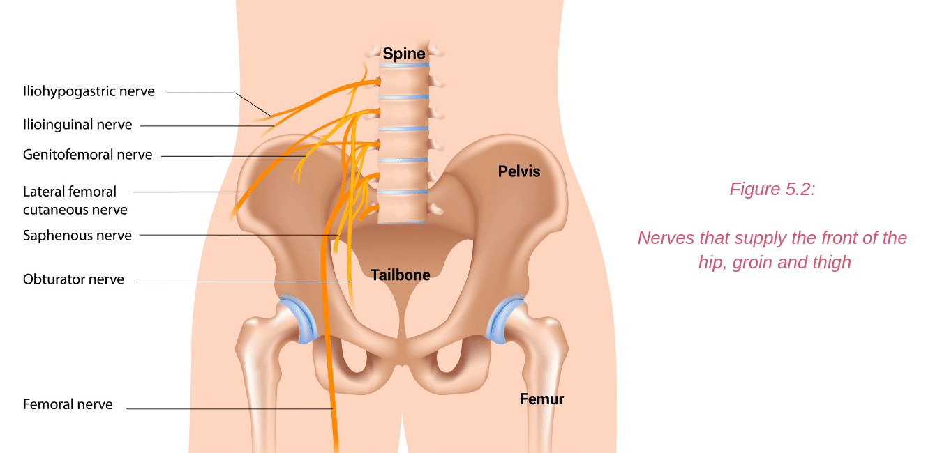
Nerves that supply the front of the hip, groin and thigh (Figure 5.2) include:
- the iliohypogastric nerve
- the ilioinguinal nerve
- the genitofemoral nerve
- the obturator nerve
- the femoral nerve
Nerve Related Pain/Neuralgia in the Anterior Hip & Groin Regions
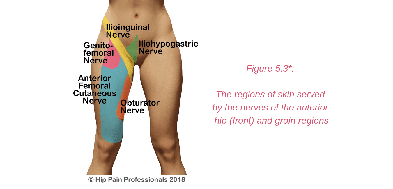
Irritation or damage to the ilioinguinal, iliohypogastric and genitofemoral nerves may occur as they travel through the muscles of the back and abdomen. Most commonly, symptoms may arise following some sort of abdominal or groin surgery, such as hernia repair.
The nerves may be damaged at the time of surgery or become entrapped in the scar tissue or mesh used for hernia repair. Endometriosis may also sometimes affect these nerves.
The symptoms are usually pain, with or without tingling or numbness in the area of nerve supply (Figure 5.3). The iliohypogastric and ilioinguinal nerves also provide some motor supply (the ability to make the muscles contract and work) to the abdominal muscles, so there may be some weakness of the abdominals experienced in conjunction with the nerve irritation.
The obturator nerve runs through the pelvic cavity and may be affected by pregnancy or pelvic surgery, as it exits the pelvis through a tunnel called the obturator canal, or as it runs between some of the deep muscles (the obturator externus & pectineus).
People with obturator neuralgia may experience pain, tingling or numbness in its area of skin supply (Figure 5.3) and may experience some weakness of the adductor (inner thigh) muscles it supplies.
The symptoms are often only felt during/after activity when the nerve has been compressed between contracting muscles.
Neuralgia of the femoral nerve is not very common, but may develop following pelvic surgery (gynaecological, prostate, bladder surgery), hip surgery or sometimes associated with problems of the adjacent soft tissue structures (psoas bursa or iliopsoasmuscle and tendon).
Femoral nerve related symptoms may be felt in the front of the thigh (in the region of the anterior femoral nerve branches that serve the skin) (Figure 5.3).
Sometimes symptoms may also be felt over the inner surface of the calf and shin (served by another branch of the femoral nerve called the saphenous nerve).
If the femoral nerve is more severely affected, muscle weakness may occur in the hip flexors (muscles at the front of the hip that lift the thigh) and the quadriceps (muscles at the front of the thigh that straighten the knee).
Nerves of the Lateral Hip Region (Side):
Nerves that supply the outer side of the hip and thigh (Figure 5.4) include:
- the ilioinguinal nerve
- the posterior branch of the iliohypogastric nerve
- the lateral cluneal nerves
- the lateral femoral cutaneous nerve
- the gluteal nerves don’t have a skin supply but can give a deep cramping feel in the buttock. They supply the gluteal muscles ability to function
Neuralgia related to the posterior branch of the iliohypogastric nerve most commonly develops due to excessive compression of the nerve as it crosses over the bony edge of the top of the pelvis to travel into the side of the hip. Here it may be affected by overly tight jeans or trousers, or from a fall onto this region, such as a fall off a bicycle or motorbike. Pain is felt in the area of its skin supply (Figure 5.5).
The lateral femoral cutaneous nerve may be compressed between the hip flexor muscles as it exits the pelvis just inside the bony point at the front of the hip (anterior superior iliac spine or ASIS).
It may also be compressed by tight belt/jeans/trousers or a large abdomen sitting down over the front of the hip (which is also more likely during pregnancy).
This nerve may also be stretched or damaged during an anterior approach Total Hip Replacement (where the scar is at the front of the hip). Symptoms developing after hip replacement surgery usually diminish or disappear over time.
Symptoms present in the outer thigh region that the nerve supplies (Figure 5.5) and are only sensory, with no impact on muscle function.
The gluteal nerves do not have a sensory supply to the skin, but gluteal neuralgia may be felt as a deep buttock pain, sometimes like a cramping feeling.
These nerves provide important motor supply to the gluteal (buttock) muscles and the tensor fascia lata (TFL) muscle at the side of the hip.
Damage to these nerves may alter your ability to stand on one leg, walk without a limp, climb stairs, and lift the leg out to the side or behind you. The nerves may be irritated or compressed as they pass out into the back of the pelvis and run through the soft tissues of the buttock.
Very occasionally, these nerves may also be damaged by surgery, such as a posterior approach Total Hip Replacement (where the scar is at the back of the hip).
Nerves of the Upper & Lower Buttock Regions
Nerves that pass through or supply the buttock region (Figure 5.6) include:
- the sciatic nerve
- the cluneal nerves–superior, middle and inferior
- the gluteal nerves –these are motor nerves that serve muscles and not the skin.
- superior gluteal nerve –serves the gluteus medius, minimus and tensor fascia lata (TFL) muscles
- inferior gluteal nerve –serves the gluteus maximus muscle
- posterior femoral cutaneous nerve–provides nerve supply to a large area of skin of the back of the thigh
Nerve Related Pain/Neuralgia in the Buttock Regions
The Sciatic Nerve and “Sciatica”
The term “sciatica”is often used incorrectly in reference to any pain felt in the area running from the back, down into the leg.
See our section on “back-related hip pain” to read more about radicular pain associated with irritation of the nerve roots as they exit the spine.
The sciatic nerve does not exit the spine as a single nerve. Nerve roots from the lower levels of the lumbar spine (lower back) and sacrum (tailbone) join together in the pelvis. Here they form a thick, cord-like structure, called“the sciatic nerve”.
This large nerve exits the inner pelvis via the greater sciatic notch and runs through the buttock and down the back of the thigh (Figure 5.7).
The sciatic nerve can sometimes be compressed, irritated or entrapped as it runs through the soft tissues of the buttock. Traditionally, sciatic pain (neuralgia) generated from issues within the buttock has been termed “Piriformis Syndrome” (see Figure 5.7 to view the piriformis muscle and the sciatic nerve).
This was based on a finding that in about 20% of the population, all or part of the sciatic nerve runs through the piriformis muscle. Compression of the nerve within the piriformis muscle was thought to be the problem in all cases of nerve related buttock and leg pain that could not be associated with a problem in the back.
It is now thought that this is the case in only a relatively small number of cases and that this condition has been over-diagnosed. So much so, that some believe it does not exist at all.
The term “Deep Gluteal Syndrome”has been suggested recently as an alternative term to piriformis syndrome. It refers to any irritation of the sciatic nerve in the deep gluteal space, beneath the gluteus maximus muscle.
In this space, the sciatic nerve may be compressed or irritated at the level of the piriformis, as it runs over the deep external rotator muscles or by fibrous bands anywhere along its path through the buttock. The nerve can also be irritated as it leaves the pelvis to head down into the thigh.
Here it runs through a tunnel (ischial tunnel), between the outer side of the sitting bone (ischial tuberosity) and the upper thigh bone (femur) (Figure 5.7).
In this tunnel it may be squeezed between the bones or irritated by unhealthy hamstring tendons (tendinopathy).
Cluneal Nerve Neuralgia
Of the cluneal nerves, the superior and inferior are more likely to be at risk of compression.The superior cluneal nerve branches run from the spine, over the top of the back of the pelvis and down into the buttock.
They usually run through fibrous tunnels as they cross the top edge of the pelvis.This is where the small nerves may become compressed or irritated.
This is usually associated with a fairly localised area of pain in the upper buttock, in the region of its skin supply (Figure 5.8).
The inferior cluneal nerve branches run across the lower buttock, right over the sitting bone (ischial tuberosity). They can be compressed and irritated by a hard fall onto the bottom or sitting for prolonged periods on a hard surface, particularly if you don’t have much gluteal muscle bulk to cushion the bone.
Again, associated symptoms are usually fairly localised to the area of skin supply (Figure 5.8). Sometimes the nearby posterior femoral cutaneous nerve can also be affected. Symptoms may then extend into the back of the thigh (see Figure 5.8 for region of this nerve supply).
Gluteal Nerve Neuralgia
The gluteal nerves do not have a sensory supply to the skin, but gluteal neuralgia may be felt as a deep buttock pain, sometimes like a cramping feeling.
These nerves provide important motor supply (the ability to make the muscles work/contract) to the gluteal muscles and the Tensor Fascia Lata (TFL) muscle at the side of the hip.
Damage to these nerves may alter your ability to stand on one leg, walk without a limp, climb stairs, and lift the leg out to the side or behind you.
The nerves may be irritated or compressed as they pass out into the back of the pelvis and run through the soft tissues of the buttock.
Very occasionally, these nerves may also be damaged by surgery, such as a posterior approach Total Hip Replacement (where the scar is at the back of the hip).
Nerves of the Saddle Region:
Nerves that supply the saddle region (Figure 5.9 and 5.10) include:
- the genitofemoral nerve
- the pudendal nerve and its branches
- the inferior cluneal nerve
- the coccygeal plexus–small nerves that supply the area around the coccyx (the bone at the very bottom of your tailbone). The anococcygeal nerve serves the skin in this area.
Nerve Related Pain/Neuralgia in the Saddle Region
Pudendalneuralgiais the most common type of neuralgia reported in the saddle region.
This small nerve may be compressed or irritated on its pathway through the buttock, where it travels from inside the pelvis to outside the pelvis and then turns around and travels into the perineal (pelvic floor) area.
It runs between the strong pelvic ligaments (sacrospinous and sacrotuberous) and through a fibrous tunnel (Alcocks canal) and may be compressed or irritated on this journey.
Unfortunately, pudendal neuralgiais often misdiagnosed.
Symptoms of pudendal neuralgia include:
- pain over the external genitalia and the skin around the anus and perineum (Figure 5.9 and 5.10)
- aggravated by sitting – sitting can put pressure on the nerve as it passes through the deep buttock area. In contrast, sitting on the toilet seat is not painful.
- pain that builds during the day but is usually better at night or when lying down, when the gravitational pressure on the pelvic floor, and thus the nerve, is reduced
- symptoms related to the nerve’s motor supply (the ability to make the muscles work/contract) to some of the pelvic muscles.Examples of this include:
- symptoms such as bladder frequency (needing to urinate frequently or urgently)
- the sensation of an urgent need to open the bowels and sexual dysfunction
- altered sensation/feelingor numbness in the perineal region (area between the pubic bone at the front, and the coccyxor tailbone at the back) served by the nerve (Figure 5.9 and 5.10)
This condition may occur:
- following childbirth or pelvic trauma or surgery
- in association with sacroiliac joint problems
- due to hypertrophy (large size) of the piriformis muscle, where the muscle presses the nerve against the overlying sacrospinous ligament
- due to direct pressure in the area – e.g. long durations cycling or during hip arthroscopy surgery where a stabilising ‘post’ may press against the perineum
Your Hip Pain Professional can:
- perform specific tests in the clinic to see if nerve involvement is likely
- provide treatments and give you exercises that may improve the health or movement of the nerve
- help improve health of the muscles and tendons beside the nerve (this may be the source of nerve irritation)
- review the positions you spend time in and activities you perform daily and provide strategies when performing these tasks that might help protect the nerve, thus reducing your symptoms. This may include changing your sitting or lying posture, or changing stretches or strength exercises that you have been performing that may be contributing to the irritation the nerve
- provide nerve gliding or mobility exercises that can be useful in some situations
- refer you for further tests or to a neurologist, orthopedic specialist or other pain specialist if required.
- In some cases, your hip pain professional may refer you to a pelvic floor physio for further assessment should they consider the pelvic floor muscles are involved.* Please note: Nerve supply can overlap and be quite variable between individuals. The diagrams provided in this section only provide an approximate guide of nerve supply in each region.
Other causes of hip, pelvic & groin pain
There are other processes that may produce pain around the hip and pelvis.
These include:
- Systemic/Rheumatological conditions: eg Rheumatoid Arthritis, Polymyalgia, Polyarthralgia
- Infective processes: Septic arthritis, Osteomyelitis, Viral Arthritis
- Neoplastic processes (cancer)
- Vascular issues: Avascular Necrosis, External Iliac Artery Entrapment, varicotic gluteal vessels compressing the sciatic nerve
These conditions are relatively rare compared to musculoskeletal pain, but when present you will need to see a medical practitioner and usually a specialist for that system or problem, for example, a Rheumatologist, Orthopaedic Specialist, Oncologistor Vascular Specialist.
Lower abdominal and pelvic pain can also be associated with organ problems (ovaries, uterus, bowel, bladder, prostate). One of the most common causes of organ-related pain in females is endometriosis.
This occurs when tissue that is similar to the lining of the uterus (womb) grows outside the uterus attaching to other structures such as the pelvic ligaments or the bladder and bowel.
This can lead to debilitating pain that may be cyclic, relating to the menstrual cycle.
Diagnosis and management of organ related problems will usually require the assistance of a specialist, for example a gynaecologist (female reproductive organs), urologist (bladder and prostate) or gastroenterologist (gut/bowel).
A skilled assessment by a Hip Pain Professional will help clarify if the problem requires further medical attention and whether it is likely to be related to a musculoskeletal problem or not.
Your Hip Pain Professional can:
- provide a detailed assessment
- assist in determining if the problem is musculoskeletal or not
- develop a plan to help if the problem is musculoskeletal
- recommend further tests or send you to another specialist if the problem does not appear to be in the musculoskeletal system.